Anatomy of the Heart
Understanding the anatomy of the heart helps you understand AFib and its complications.
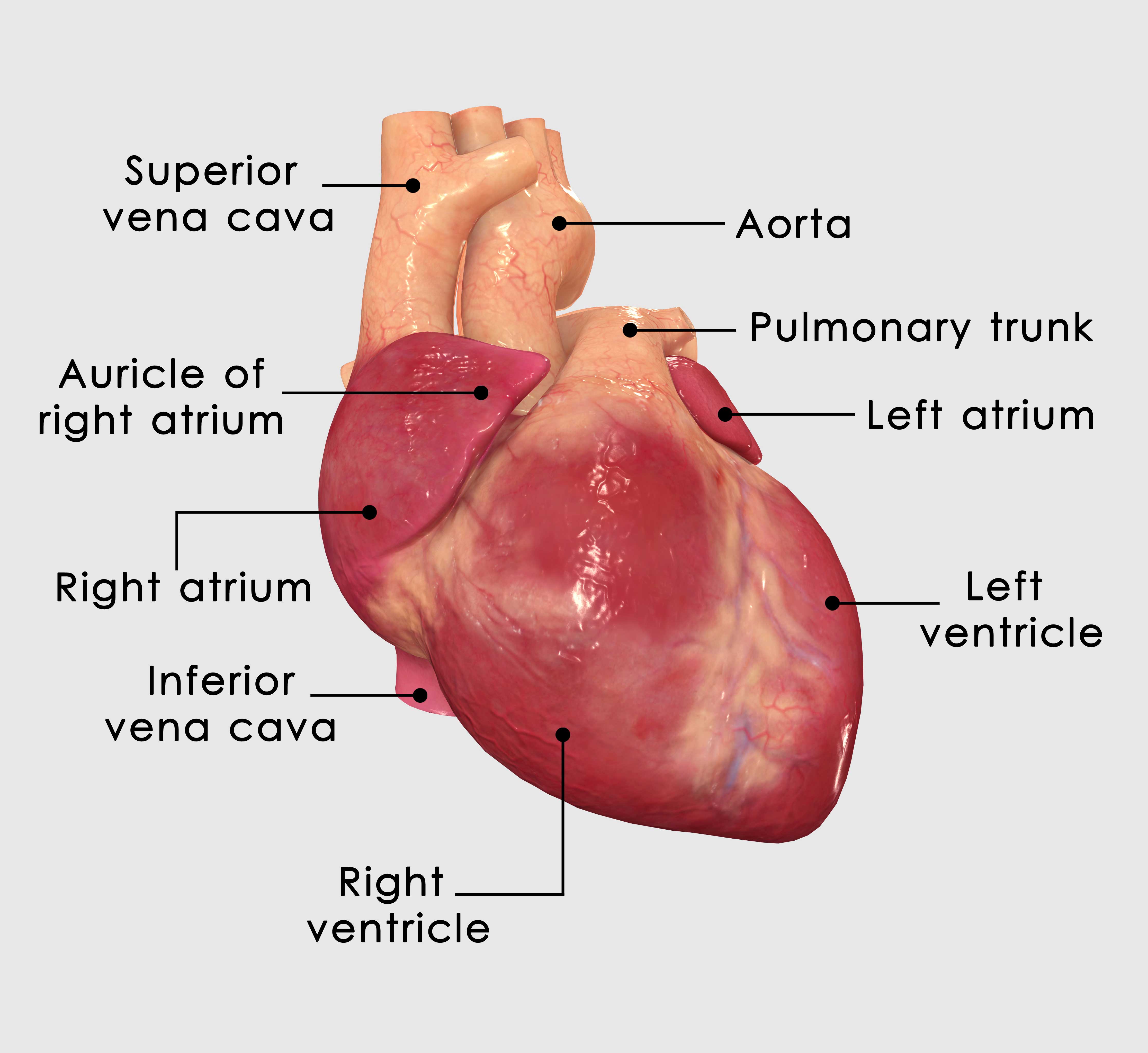
The heart has two upper chambers called the right atrium and left atrium. The two bottom chambers are called the right ventricle and left ventricle.
The bottom heart chambers are big, thick, and muscular, and these serve as the main pumping chambers for the heart. All the veins in the body return the deoxygenated blood to the heart through the superior and inferior vena cava into the right atrium. The blood then flows through a valve into the right ventricle which is the main pump that sends the blood to the lungs to get oxygen. The blood then returns to the left atrium, flows through another valve into the left ventricle, and then the left ventricle acts as the main pump out to the body to supply it with blood. This is the basic plumbing of the heart.
The electrical system starts in the upper right corner of the right atrium with a structure called the sinus node or SA node. This is the heart’s own pacemaker that all people are born with. It functions to regulate the heart rate which is typically between 60 to 100 beats a minute at rest. The sinus node sends signals out in a very coordinated/regular fashion, creating the normal heart rhythm. The top and bottom chambers are electrically separate from each other except for a single connection in the center of the heart called the AV node. This structure sends the signal from the upper chambers to the bottom chambers (left and right ventricles).
AFib in the Heart
AFib is an abnormal heart rhythm that is not generated from the sinus node in the right atrium but rather originates from the left atrium. Tissue in the left atrium, most commonly from the pulmonary veins (which are the veins that return blood from the lungs to the left atrium), sends out its own electrical signals to stimulate the heart to beat.
These are the signals that trigger AFib. Unlike normal rhythm, the AFib signals are chaotic and irregular, beating much faster at nearly 600 beats a min in the upper chambers. Not all of these signals are transmitted to the lower chambers (the ventricles) which are the main pumps to move blood through the body. This results in the upper and lower chambers no longer working together synchronously. It is this chaotic, asynchronous rhythm that affects how the normal ventricles pump and leads to the typical symptoms of AFib.
Unknown Cause
The underlying cause of AFib remains unknown despite the fact that it is very common in the general population. Unfortunately, AFib is a progressive disease. It typically begins with short episodes interspersed between long periods of normal heart rhythm. This is known as paroxysmal AFib. Over time, these episodes begin to occur more frequently and last longer in duration. This is known as persistent AFib. If left untreated, some patients will progress to being in AFib continuously without any periods of normal rhythm. Eventually, this can become permanent AFib.
Early identification and treatment of AFib are key to maintaining normal heart rhythm.